Tethered Spinal Cord
Key Points
- Bones are lifted up (Laminoplasty) rather than removed (Laminectomy) to perform most tethered cord surgery
- Nerves to the legs and bladder are always monitored and protected during surgery
- Laser is used to remove Lipomas. (see a video of the procedure)
What is Tethered Cord?
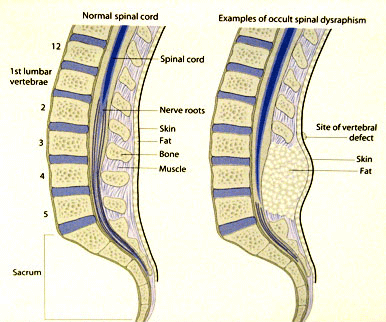
The spinal cord includes the bundle of nerves that controls leg movement and sensation as well as bladder function. The spinal cord typically divides into small nerve roots at the L2 vertebral body. During development of the spinal cord, tissue and fat, or other body elements that do not belong near the spinal cord can become attached to the spinal cord. Sometimes the tissue prevents the normal development of the spinal cord so that there are problems with urination and leg weakness. In most cases, there are no problems at birth. As the body grows, however, the spinal cord then becomes stretched and damaged by the abdominal attachment. This condition is called a tethered spinal cord. If left untreated, your child may suffer nerve damage as he or she grows. The condition can be treated with surgery to prevent future nerve damage.
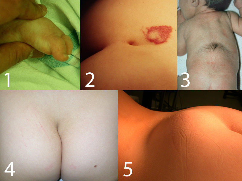
Signs and symptoms of a tethered cord can include the following:
- A crooked toe. (Photo 1)
- A dimple above the gluteal crease (the crease in the buttocks) (Photo 2)
- Long hair (longer than 1 inch) growing on the back over the spine. (Photo 3)
- A crooked crease between the buttocks. (Photo 4)
- A lump of the lower back. (Photo 5)
- Stumbling or changes in gait or walking.
- Pain or tingling the legs or back
- Curvature of the spine
- Trouble with bowel or bladder control, such as difficulty in toilet training in a toddler, keeping a dry diaper with a baby, loosing control in a toilet trained child or no being able to hold urine until getting to the bathroom.
The diagnosis of a tethered spinal cord is made by obtaining a Magnetic Resonance Imaging or MRI of the spine.
Surgery for Tethered Cord
Surgery involves the following steps:
- Needles are inserted into the lower body to monitor nerve function even as the child is asleep.
- An incision is made in the skin over the lower back
- Lifting up of the bone over the tethered area of the spinal cord ( Laminoplasty)
- Making a small opening in the covering of the spinal cord, called the dura
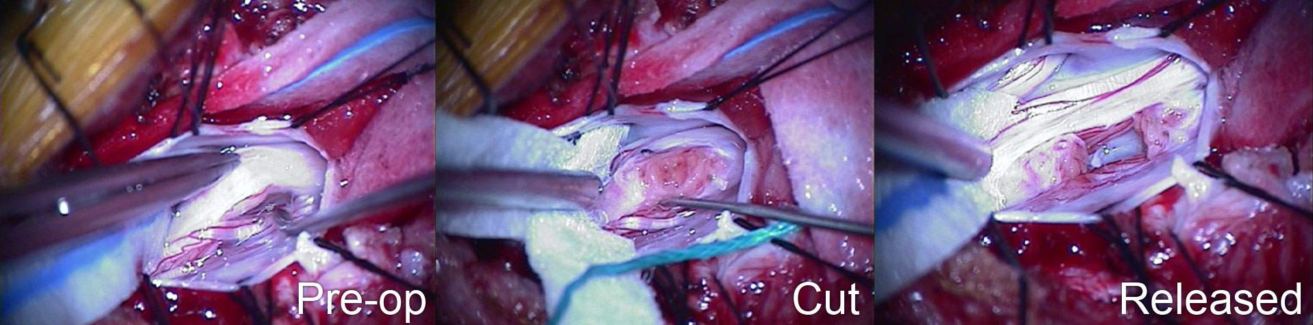
- Releasing the tethering lesion; can be simple or highly complex requiring many hours or dissection under the microscope. At times we use a laser to sharply dissect tissue from the nerves. The images below show the lesion before it is cut (left), the lesion after it is cut (center), and the lesion released (right).
- Closing the dura or available tissue over the spinal fluid.
- Replacing and securing the bone (laminoplasty movie coming soon)
- Closing the muscle and skin
- Applying a bandage
Surgery for a tethered spinal cord can prevent future irreversible nerve damage which can severely affect a child's bladder and leg function and quality of life.
Re-Operation for Tethered Cord
A follow-up MRI is usually performed sometime after surgery. On rare occasions, years after the original surgery, a re-tether may develop for certain types of tethered cords causing radiating leg pain, persistent back pain or bladder problems. This situation may require additional surgery.